


dSTORM image of the tubulin cytoskeleton of U2OS cells where the depth of the tubulin fibers along the vertical axis was resolved by using astigmatism in the detection optics introduced by the addition of a cylindrical lens.
General description of single molecule localization microscopy (SMLM) techniques
The potential of utilizing single molecule fluorescence for achieving optical superresolution was initially postulated in a short and somewhat cryptic paper by Betzig, published in 1995. It took until the year 2006, before three research groups independently demonstrated similar optical reconstruction methods with a lateral sub-diffraction optical resolution of better than 20 nm. While Eric Betzig and collaborators, now with the Howard Hughes Medical Institute at the HHMI Janelia Farm Research Campus, termed this method “Photoactivated Localization Microscopy” (PALM), S. T. Hess and colleagues from the University of Maine called it “Fluorescence Photoactivation Localization Microscopy” (FPALM).
The importance of breaking the diffraction limit is demonstrated by the award of the Nobel Prize in Chemistry 2014 to Eric Betzig (UC Berkeley, LBNL, and HHMI Janelia Research Campus), Stefan W. Hell (MPI for Biophysical Chemistry and University of Göttingen; German Cancer Research Center, MPI for Medical Research, and University of Heidelberg) and W.E. Moerner (Stanford University) for the development of super-resolved fluorescence microscopy and the recent Breakthrough Prize in Life Sciences 2019 awarded to Xiaowei Zhuang for the development of Stochastic Optical Reconstruction Microscopy (STORM).
These photoswitching techniques all have in common that they exploit the temporal separation of single fluorescent emitters, even if the molecules cannot be isolated in space due to the diffraction limit: Multiple localizations of single molecules obtained in a series of images are used to reconstruct a super-resolved image based upon the positions obtained from the localization algorithm.
All of these methods overcome the problem of resolving structures with sizes below the optical diffraction limit and which consist of fluorescent molecules by utilizing special properties of photoactivatable proteins or photo-switchable fluorophores that make it possible to tell if the fluorescence emission was due to a single molecule or not. FPALM uses fluorescent proteins such as the photoactivatable green fluorescent protein (GFP), while PALM also makes use of photoswitchable proteins. The first fluorophores that were used for STORM were carbocyanine fluorophores, which exhibit photoswitching properties in the presence or absence of an activator molecule. The latter method, where the bare fluorophore is used in a special buffer system, is therefore called directSTORM (dSTORM)1,2. Fluorophores are either switched between a fluorescent bright (“on”) and a non-fluorescent dark (“off”) state upon illumination with light of different wavelengths (PALM/STORM) or they are photoactivated and subsequently photobleached (FPALM).
Due to their technically relatively simple implementation, wide-field single-molecule based localization approaches such as PALM, STORM, and dSTORM are currently widely used for super-resolution imaging. It is even possible to gain an improved axial resolution by introducing astigmatism due to a cylindrical lens into the detection optics or by shaping the detected point spread function (PSF) of a single emitter with adaptive optics. The distortions that this causes to the PSF can be characterized and enables one to gain a super-resolved image with up to several microns depth. Further applications of photoswitching microscopy are comprised of studying slow dynamics in living cells anchored to a substrate and quantitative high-resolution fluorescence imaging, e.g. studies that count the number of biomolecules and their structural organization in small subcellular structures, e.g. organelles or the cell membrane. Super-resolution optical microscopy techniques are also attracting attention in the fields of chemistry and material science, e.g. by resolving the morphology of microgel particles via dSTORM measurements.

The images to the left (a, d and g), are transmission light micrographs of NIPAM based microgels synthesized with 5.0 mol%, 7.5 mol%, 10.0 mol% BIS (from left to right). The middle row shows diffraction limited fluorescence images summed up from the 50 000 single frames acquired for the dSTORM reconstruction (b, e and h). Comparison between the white light and the fluorescence images shows that the main part of the fluorescence signal has its origin located on the inside of the microgels. The number of localizations is encoded in the color-coded grey values of the reconstructed images (c, f and i) by applying the red hot look up table. The number of localizations is found to increase with growing cross-linker content during the synthesis. Image size is 15.87µm x 15.87µm. Scale bar 1µm
The next generation: dSTORM II System
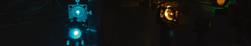
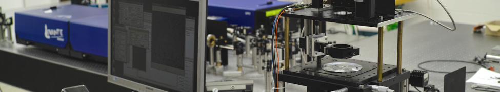
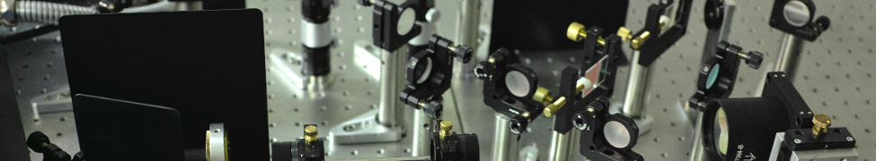
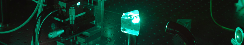
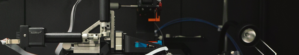
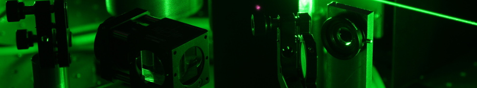
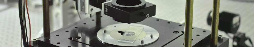
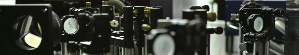
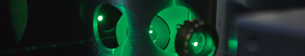
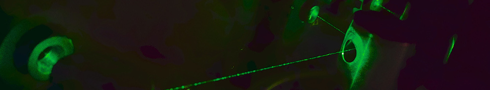
Our dSTORM setup II is based on an Olympus IX71 widefield microscope with an Argon-Krypton ion laser source which enables us to excite several different fluorescent proteins and organic fluorophores. Additionally, a 405 nm diode laser is available to recover organic fluorophores to the “on”-state to control the emitter density during data acquisition. If the emission of the 405 nm diode laser is controlled by a function generator, then this enables us to conduct classic photoactivation localization microscopy (PALM) by pulsed activation with subsequent localization and photobleaching. Based on the recent publication by Diekmann et al.3 , 2 low cost industry grade CMOS cameras are used in this setup. Different illumination schemes, from epi-illumiation over HILO to TIRF, are implemented using a 60x TIRF oil immersion objective lens.
In comparison to our dSTORM setup I, which still uses electron-multiplying CCD cameras and is also outfitted with an optical tweezers system, the dSTORM II setup also enables us to acquire two color channels simultaneously, where one emission channel is reserved for an emission wavelength >650 nm, typically Alexa Fluor 647. Based on the approach to gain an improved axial resolution by introducing astigmatism with a cylindrical lens it is further possible to perform 2 color 3D dSTORM measurements.
A future development of the setup will be the introduction of a deformable mirror to apply adaptive optics methods, such as corrections of spherical aberrations or even to obtain the possibility to localize molecules along the vertical axis at a depth of up to 5 microns4 via PSF engineering.
Recent applications of SMLM in our research group
Beside the application of SMLM to biological samples, SMLM can also be applied to polymer science and material science.
Since 2016 SMLM gains more attention in the field of polymer science since interesting properties such as the response of external stimuli, e.g. temperature, electrical fields or pH value, can be measured dynamically and in-situ avoiding the problems and limitations associated with sample preparation for electron microscopy. By localizing unbound rhodamine 6G within the polymer particles it is possible to resolve the network morphology5.
We also utilize dSTORM based on optical waveguide illumination6 ("Waveguide-based optical nanoscopy"), as well as in nonadherent cells by combination with optical tweezers7.
Together with the research group of Prof.Dr. H. Milting from the Erich and Hanna Klessmann Institute for Cardiovascular Research & Development at the Heart and Diabetes Centre NRW we tackle the question of the interaction between different mutations of proteins located in human heart cells (cardiomyocytes) responsible for the proper contraction of the human heart.

Photoactivation Localization Microscopy (PALM) reconstructions of mEos2 (red fluorescence) fused to Plakophilin-2 and merged with white light transmission images of the cells. The wild-type Plakophilin-2 mEos2 signals is localized at the plasma membrane (a). Removing the tail domain has no visible effect on the localization position (b). Without Armadillo repeats Plakophilin-2 is not specifically localized at the membrane (c). Scale bar 10 µm.
References
(1) Wolter, S.; Schüttpelz, M.; Tscherepanow, M.; van de Linde, S.; Heilemann, M.; Sauer, M. Real-time computation of subdiffraction-resolution fluorescence images. Journal of microscopy 2010, 237, 12–22, DOI: 10.1111/j.1365-2818.2009.03287.x.
(2) Heilemann, M.; van de Linde, S.; Schüttpelz, M.; Kasper, R.; Seefeldt, B.; Mukherjee, A.; Tinnefeld, P.; Sauer, M. Subdiffraction-resolution fluorescence imaging with conventional fluorescent probes. Angewandte Chemie (International ed. in English) 2008, 47, 6172–6176, DOI: 10.1002/anie.200802376.
(3) Diekmann, R.; Till, K.; Müller, M.; Simonis, M.; Schüttpelz, M.; Huser, T. Characterization of an industry-grade CMOS camera well suited for single molecule localization microscopy - high performance super-resolution at low cost. Scientific reports 2017, 7, 14425, DOI: 10.1038/s41598-017-14762-6.
(4) Aristov, A.; Lelandais, B.; Rensen, E.; Zimmer, C. ZOLA-3D allows flexible 3D localization microscopy over an adjustable axial range. Nature communications, 9, 2409, DOI: 10.1038/s41467-018-04709-4.
(5) Bergmann, S.; Wrede, O.; Huser, T.; Hellweg, T. Super-resolution optical microscopy resolves network morphology of smart colloidal microgels. Physical chemistry chemical physics : PCCP 2018, 20, 5074–5083, DOI: 10.1039/c7cp07648g.
(6) R. Diekmann, O.I. Helle, Cristina I. Øie, P. McCourt, T.R. Huser, M. Schüttpelz, and B.S. Ahluwalia, Chip-based wide field-of-view nanoscopy, Nature Photon. 11, 322-328 (2017)
(7) R. Diekmann, D.L. Wolfson, C. Spahn, M. Heilemann, M. Schüttpelz, and T. Huser, Nanoscopy of bacterial cells immobilized by holographic optical tweezers, Nature Commun. 7, 13711 (2016), DOI: 10.1038/ncomms13711