|
|
|
|
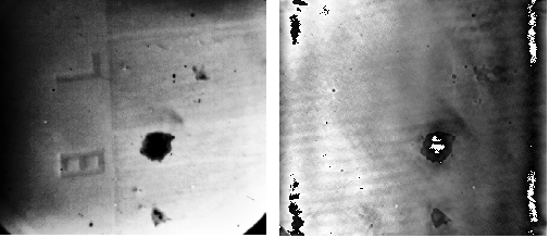
Our main research on photoelectron microscopy in the EUV and SXR spectral range is focused on the characterization of nanostructured surfaces and buried interfaces of ultrathin organic and metallic nanolayers. In contrast to UV excitation where work function differences and topographic effects dominate the image contrast, excitation with higher photon energies reach the electron core levels of many materials (e.g. the 3p levels of Ti, V, Cr, Mn, Fe, Co, Ni, Cu, Zn). Besides topographic imaging a chemical contrast can be achieved by tuning the photon energy to X-Ray absorption edges in the EUV and SXR spectrum. The technique allows the study of interdiffusion and interface reaction processes insitu with a lateral resolution of <100 nm.
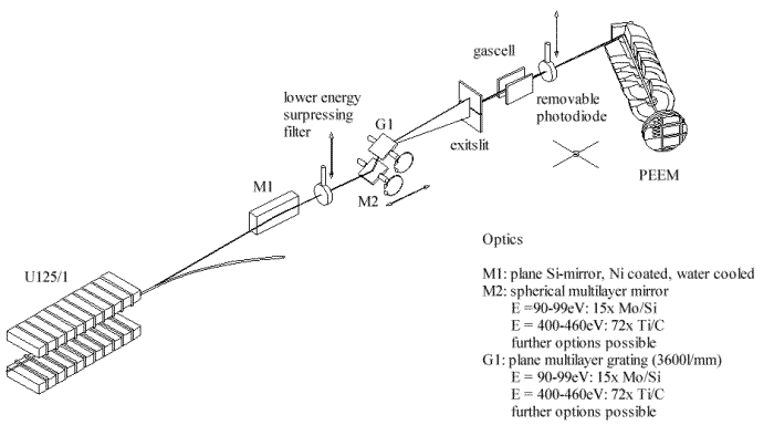
A high flux multilayer monochromator for the photon energy ranges 90 to 99 eV and 400 to 460 eV was developed by using two interchangeable sets of focusing multilayer mirrors and multilayer gratings. These optical elements have been coated by our EUV optics group. An extension to photon energies in between 100 eV and 400 eV using of other optical multilayer elements is also possible.
The current development of a specially developed "wet cell" in transmission geometry will allow imaging of biomaterials (cells, lipid films) in a hydrated state in the "water window" spectrum (280 eV - 500 eV) in the near future.
"Photoemission Microscopy with Microspot-XPS by use of Undulator Radiation and a High-Throughput Multilayer Monochromator at BESSY"
U. Kleineberg, D. Menke, F. Hamelmann, U. Heinzmann, O. Schmidt, G.H. Fecher, G. Schönhense
J.Elect.Spect.Rel.Phen . 101-103 (1999) pp. 931-936
"Time-of-flight photoelectron emission microscopy TOF-PEEM : First results"
H. Spiecker, O. Schmidt, Ch. Ziethen, D. Menke, U. Kleineberg, R.C. Ahuja, M. Merkel, U. Heinzmann, G. Schönhense
Nucl. Instrum. Meth. A 406 (3), pp. 499-506 (1998)
"Chemical microanalysis by selected-area ESCA using an electron energy filter in a photoemission microscope"
O. Schmidt, Ch. Ziethen, G.H. Fecher, M. Merkel, M. Escher, D. Menke, U. Kleineberg, U. Heinzmann, G. Schönhense
J. Electr. Spectr. Rel. Phenom . 88-91 , pp. 1009-1014 (1998)


|
Text von
Martin Pohl , Ulf Kleineberg
|
Letzte Änderung
03.07.2001 |
| Wir haben auf unseren Seiten Hyperlinks zu anderen Seiten im Internet gelegt, deren Webmaster wir nicht sind. Für alle diese Hyperlinks gilt: Wir erklären hiermit ausdrücklich, dass wir keinerlei Einfluss auf die Gestaltung und Inhalte dieser Seiten haben. Deshalb "distanzieren" wir uns hiermit von allen Inhalten dieser Seiten und machen uns ihre Inhalte in keiner Weise zu Eigen. | ||
|
©2001
Molekül- und Oberflächenphysik
Universität Bielefeld |
||