| |
 |
Biophotonics of Cells & Tissue
- CELL IMAGING
Contact: Dr. Katja Tönsing |
| |
|
|
| |
|
Living biological cells and tissue are being
imaged and investigated with respect to their cell surface and
global internal structure via in-situ atomic force microscopy
(see paragraph AFM), confocal optical (2-photon) laser scanning
microscopy and TIRF microscopy (see paragraph Single Molecule
Photonics).
For optical cell imaging we currently use a single- or multifocal
(1-64) 2-photon laser scanning microscope driven by a 1,5 Watt
Ti:Sa fs-laser (adjustable to 700-960nm) for confocal 2-photon
excitation and detection. The three main advantages of this
instruments are a) the focal excitation volume compares favorably
with conventional 1-photon excitation and allows distinct “optical
point like” excitation and activation, b) the excitation
wavelength in the near IR gives less rise to unwanted light
scattering phenomena in biological cells and tissues, and c)
the 2-photon wavelength can be adjusted to the autofluorescence
properties of proteins (e.g. collagen), and to the protein marker
GFP and GFP-analoga, respectively. |
| |
|
|
| |
|
 |
| |
|
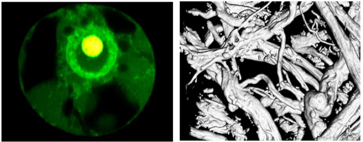
Images: 2Ph-LSM of GFP-expression in living
plant cell and of collagen tissue |
| |
|
|
| |
Project 1 |
GFP Expression in Living Cells |
| |
|
The distribution and dynamics of GFP expression
and protein translocation in living plant cells is investigated
by 2-photon laser scanning microscopy (see image). This result
is a first step to investigate the distribution and dynamics
of fluorescently labelled GTP, G-proteins, G-protein receptors
and photoactivatable proteins within a cellular organism. |
| |
|
Publications: [see section "Publications"] |
| |
|
Collaboration: T. Merkle and K. Niehaus, Dept.
of Biology, Bielefeld University |
| |
|
|
| |
Project 2 |
Biomedical Characterization of Collagen
Tissue for Autologous Tissue Transplantation |
| |
|
The characterization collagen grafts with
chondrocytes for Matrix-induced autologous chondrocyte implantation
(MACI) is crucial for biomedical applications in tissue engineering.
The simultaneous imaging of the collagen matrix and the chondrocytic
activity is very difficult in this strongly scattering tissue
material, however, possible with 2-photon CLSM with remarkable
penetration depth of up to 200-500 µm. |
| |
|
Publications: [see section "Publications"] |
| |
|
Collaborations: IBA-Heiligenstadt, Fa. Verigen,
Leverkusen; Fa. Lavision-Biotec, Bielefeld |
| |
|
|
| |
Project 3 |
Biomedical Characterization of Human Cartilage
Tissue |
| |
|
Biomedical characterization of healthy and
artritic cartilage tissue with direct 2-photon LSM and non-staining
protocols. |
| |
|
Publications: [see section "Publications"] |
| |
|
Collaboration: Dr. med. M. Dickob, Bielefeld |
| |
|
|
| |
|
other ongoing projects [...] |
| |
|
|
| |
|
We acknowledge funding from BMBF Research
Initiative Biophotonics - MEMO (Germany) |